Abstract
A wearable smart insole system accurately measures foot pressure, providing a new tool for diagnosing and monitoring flat feet.
Researchers developed a portable, wearable smart insole system with flexible sensors to measure foot pressure. The system was tested on people with flat feet, healthy individuals, and those with postural deformities. The results showed that the system's pressure maps matched those of a gold-standard reference device, indicating its accuracy and usability for mobile clinical measurements.
Full TextOpen Access
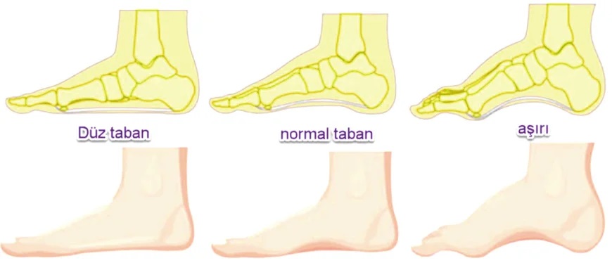
Pes planus is a condition in which the sole of the foot comes into full or near-full contact with the ground as a result of a reduction or complete loss of the medial longitudinal arch (Figure 1). Determining plantar pressure distributions and gait-cycle dynamics is a critical clinical requirement for diagnosing anomalies in lower-extremity biomechanics, evaluating patient-specific treatment protocols, and monitoring orthotic/insole designs. Although conventional pedobarography platforms provide high spatial resolution, they are inadequate for mobile follow-up because they are dependent on laboratory settings, can scale only a limited area of a few steps in the patient’s natural walking environment, and cannot fully reflect real-time stance variations during dynamic locomotion. Recent advances in wearable sensor technologies have made it possible to overcome laboratory constraints and collect continuous, natural gait data during patients’ activities of daily living [1]. Accordingly, the accessibility of practical, usable, and affordable measurement technologies that can be integrated directly into footwear for use in non-clinical settings and open-field environments without restricting freedom of movement has become a current need in biomedical engineering.
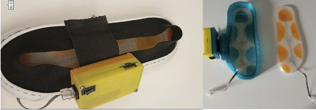
In this study, an independently operating mobile pedobarography and embedded data acquisition (datalogger) system was implemented to address the above-mentioned clinical requirements. The principal distinguishing feature of the developed device is that, without requiring an external computer connection, it timestamps the data instantaneously at a stable sampling rate of five samples per second through an integrated real-time clock module and stores the records locally in .txt format. Supported by a microcontroller architecture and a regulated battery circuit, the portable recording unit was mounted inside a compact protective enclosure made of rigid polymer material by 3D printing and clipped to the outer side of the shoe.
The flexible sensing insole component of the system was produced from Velostat material through a layered mechanical manufacturing workflow according to EU size 36 foot geometry. To eliminate the nonlinear signal noise generated by ready-made commercial force-sensitive resistor (FSR) sensors on bending surfaces, the integration of flexible Velostat polymer materials, which constitute the structural basis of commercial FSR sensors, into wearable sensor architectures was preferred [2]. Previous studies have also confirmed that flexible transducer designs successfully overcome the anatomical incompatibility and thickness-related limitations of rigid industrial sensors and provide high stability in real-time medical bracing, orthotic applications, and biomechanical pressure-monitoring processes [3]. During manufacturing, conductive pathways were printed on a flexible carrier layer using flexible conductive strips to transmit data from six critical anatomical focal points that bear the greatest vertical loads on the plantar surface: the hallux, metatarsal regions, medial arch, lateral arch, and heel. Flexible polymer-film material with resistance that changes stably according to the applied force was positioned at these focal points. To prevent signal loss due to moisture and friction, the entire insole structure was isolated by coating it with a flexible insulating protective film; subsequently, the upper and lower layers were covered with flexible polymer filament material using 3D printing to prevent shear-induced noise. To verify the sensitivity of this integrated device in discriminating anomalies, test protocols involving both static and dynamic sessions were carried out on healthy participants and patients with postural disorders. The dynamic time-series data outputs obtained from the system were compared with the outputs of a gold-standard reference clinical pedobarography device performing baropodometric measurements (Medicapteurs™), thereby validating the hardware, software, and manufacturing performance of the system in a clinical setting.
The literature has further demonstrated that Velostat-based ultra-thin flexible transducer designs overcome the anatomical mismatch and thickness constraints associated with rigid industrial FSR sensors and provide high stability in real-time medical bracing, orthotic, and biomechanical pressure-monitoring systems [5].
2.1. Needs Analysis for an Accessible and Sustainable Mobile System
The major methodological limitation of clinical pedobarography processes is that patients are made dependent on rigid, immobile, fixed floor plates in laboratory environments. This restricts natural locomotion and walking dynamics and prevents the capture of real-time stance variations. Rather than being designed around a cost-based limitation, the proposed wearable system enables the patient to generate continuous, unrestricted, and natural data while wearing their own footwear in non-clinical environments, open-field settings, and during activities of daily living. In this respect, the system was implemented as a mechanically sustainable, functionally usable, practical mobile pedobarography solution with high clinical adaptability.
2.2. Biomechanical Design and Layered Manufacturing Processes of the Original Sensing Insole
The thick and rigid protective films on the outer surfaces of commercial force sensors produce folding-induced noise on continuously bending and mobile surfaces such as the inside of a shoe, thereby impairing data stability. To eliminate these nonlinear instabilities and data losses, ready-made sensor architectures were completely abandoned. The hardware philosophy of the system is based on adapting a 100 μm-thick piezoresistive Velostat polymer-film material, which undergoes molecular compression under vertical pressure and whose electrical resistance changes inversely with the applied force, into an ultra-thin and flexible form suitable for the constraints of in-shoe use. The electrical conduction mechanisms of such polymeric matrix structures under vertical loading have been characterized in the literature by percolation theory and quantum tunneling effects between conductive carbon particles [6].
The flexible sensing matrix of the system was brought to its final ergonomic form through a staged mechanical production line following four fundamental research-and-development steps:
Anatomical Localization and Printing of Conductive Traces: During manufacturing, six critical anatomical regions where vertical ground-reaction forces are most concentrated throughout the gait cycle were selected as focal points. These focal points were the hallux, first metatarsal, third metatarsal, medial longitudinal arch, lateral longitudinal arch, and calcaneus (heel). On the high-strength and highly flexible carrier-film layer forming the main body of the insole, the traces that would transmit data from these focal points to the microcontroller were processed with millimetric precision using flexible conductive strips.
Sandwich Architectural Configuration and Insulation: A 100 μm-thick piezoresistive polymer-film material was placed between two oppositely positioned conductive-strip layers. To prevent signal losses caused by moisture, perspiration, and friction, as well as mechanical bending noise, the entire cell structure was mechanically sealed and insulated using flexible, highly insulating protective strip films thinner than 50 μm.
Flexible Additive-Manufacturing Integration: To prevent the manufactured flexible sensor nodes from shifting during in-shoe mobility and to ensure that they received vertical forces at a fully perpendicular angle, the lower and upper outer protective layers of the insole were printed from flexible filament material using 3D printing (additive manufacturing) technology, and the sensor matrix was permanently embedded into this flexible base during production.
Producing all six circular/oval flexible sensing cells with identical surface-area dimensions, a common geometry, and the same load-resistance characteristics provided the system with a considerable mathematical advantage. Owing to this hardware standardization, the need to define a separate mathematical correction coefficient for each sensor node at the microcontroller level was eliminated, and a homogeneous and stable data flow across the entire insole was achieved through a single shared calibration model.
2.3. Standalone Portable Datalogger Unit and Mechanical Enclosure
To record data without restricting the patient’s freedom of movement and without the need for an external computer connection, a fully standalone “black-box” datalogger architecture was designed.
Central Processing Unit and Power Management: At the core of the hardware backbone of the system, a central microcontroller unit was positioned because of its low power consumption, compact dimensions, stable interrupt management, and the fact that its architectural design limits and electrical pin map are documented in industrial datasheets. To enable the system to operate independently in mobile environments, a 3.7 V rechargeable battery integrated with an externally powered charging module was used. The integrated power-management layer in the system maintains the stable operating voltage required by the hardware and sensor cells, thereby ensuring homogeneous energy flow to all lines. The real-time clock integrated circuit that governs chronological tracking in the hardware architecture has leap-year-compensated calendaring capability and an ultra-low-power backup mode that preserves system integrity during instantaneous fluctuations in the main battery, as specified in the manufacturer documentation. In the storage layer of the datalogger unit, an integrated logic buffer circuit and voltage regulator were used to reduce the logic output levels of the central processor to the nominal level tolerated by the memory card. This hardware scheme prevents data corruption that may occur during block-writing sessions on the card during dynamic walking.
Mechanical Enclosure Design (Rigid Material Strength): All embedded-system components were placed inside a custom enclosure to protect them from external environmental conditions, vibration, humidity, and mechanical impacts. Additive-manufacturing technology was used in this process, and robust protective boxes were fabricated from rigid filament material with high impact resistance, structural stiffness, and dimensional stability. This mechanical interface, which can be conveniently clipped to the outer edge of the shoe or to the user’s ankle, transforms the system into an independent device that can be used beyond the boundaries of the clinical environment.
2.4. Real-Time (Current Date/Time) Data Storage and Embedded Software Filtering
The embedded software architecture was optimized to convert analog data from the sensors into digital signals and transfer them to local storage with chronological accuracy.
Real-Time Logging: Analog voltage signals varying according to plantar pressure are read through the analog input channels of the microcontroller. In dynamic gait-cycle analyses, determining the exact millisecond at which data are recorded is critical for clinical follow-up. For this purpose, step data are timestamped instantaneously with a stable sampling frequency of five samples per second (5 Hz) using the real-time clock module integrated into the system. The time-series outputs are continuously stored in local memory in the open-source .txt format by means of the storage module.
Software-Based Lower-Threshold Filtering Algorithm: Because the flexible sensor cells have high sensitivity, a software-based lower-threshold filter of 200 ADC counts was incorporated into the embedded software to remove single-point micro-contact noise and instantaneous noise spikes (artifacts) arising while participants put on the shoe, during unloaded initial contacts, or while the foot is airborne during the swing phase. All raw digital data (0-1023) below this threshold were automatically set to the baseline value (zero) by the software, thereby successfully filtering irrelevant time intervals and idle-operation noise before clinical analysis.
The hardware stability, measurement sensitivity, and biomechanical discriminative capacity of the developed portable pedobarography system, which has high usability and accessibility, were tested through clinical sessions conducted at the Gait Analysis Laboratory of Metin Sabancı Baltalimanı Bone Diseases Training and Research Hospital (Türkiye). All testing and measurement procedures were performed with reference to the laboratory’s standard clinical pedobarographic validation and data-collection protocols. To establish the clinical performance limits of the system, integrated test protocols covering static stance and active dynamic walking phases were conducted in target groups consisting of orthopedically healthy participants and patients with clinically evident stance and postural disorders.
The time-series logs that the system hardware continuously transferred to local storage were analyzed with reference to participants’ physical anthropometric characteristics, mass distributions, and anatomical foot dimensions. Instantaneous micro-contact noise and idle-operation signals caused by vertical load imbalances during the process of putting on the shoe, during unloaded initial contact, or during waiting periods were successfully removed from the dataset before clinical analysis by means of the 200 ADC software lower-threshold filter integrated into the embedded software and the applied data-preprocessing methods.
To verify the sensitivity of the system in isolating pathological vertical load accumulations and kinetic asymmetries on the plantar surface, data obtained from the patient group with stance and postural disorders were analyzed in detail. In the clinical sessions conducted under the coordination of Assoc. Prof. Dr. Osman Nuri Özyalvaç and under the supervision of a specialist physician, integrated test protocols covering static stance and active dynamic walking phases were applied to patients included in the study sample who had a marked stance anomaly. As emphasized in pediatric-patient-oriented validation studies on flexible pes planus that associate pedobarographic analysis hardware with the clinical asymmetries of radiological findings, medial displacement of vertical ground-reaction forces is one of the most decisive factors in monitoring clinical diagnostic processes [4].
When the chronological time-series data collected from six critical anatomical focal points on the insole throughout the participants’ locomotion cycle were examined, the system demonstrated full biomechanical consistency and a high degree of correlation with the patient’s existing orthopedic pathology. In a patient with a postural disorder, the center of mass of the left foot was found to shift markedly toward the medial axis during stance (pronation tendency), and vertical ground-reaction forces (vGRF) were concentrated along the medial edge of the plantar surface. During the static stance phase, extremely steady and stable signals close to the maximum measurement limit were obtained for an extended period from the sensor cell representing the left-foot calcaneus (heel) focus; simultaneously, a sustained high load accumulation was recorded in the medial metatarsal region due to mass loading. During the dynamic stepping cycle, marked increases and kinetic fluctuations were observed in the vertical-load graphs of the medial arch region as the participant’s center of mass collapsed inward. In contrast, the lateral arch focus remained considerably subdued, stable, and low-level throughout the walking phases because it could not compensate for the mass distribution. These stable and asymmetric curves produced by the developed flexible sensing transducer matrix directly on the patient confirm that the system can identify anomaly locations, load shifts, and kinetic deviations arising from postural and stance disorders in non-laboratory settings with high isolation and diagnostic success.
To verify the hardware manufacturing symmetry, calibration accuracy, and homogeneous measurement capability of the system, data obtained from the healthy control group with completely balanced anatomical structure were subjected to integrated analysis. In both static postural stance and consecutive dynamic walking phases of healthy participants, the time-series data traces obtained from the right and left insole channels were observed to follow each other morphologically in mirror symmetry. Throughout the gait cycle, the periodic transfer of vertical force beginning with heel strike and progressing toward the metatarsal foci and toe tip was temporally documented. The fact that differences between corresponding right and left sensor locations remained within a flexible symmetry band statistically close to zero during the dynamic cycle, together with the strong convergence of the right/left calibration coefficients, demonstrated with high stability that the device could measure the geometric standardization achieved in sensor-cell manufacturing and the state of full balance.
3.3. Mathematical Modeling-Based Heat Maps and Reference Validation
To convert timestamped time-series logs stored in text format on the MicroSD card into clinically scannable and interpretable visual outputs, open-source engineering algorithms and mathematical data-processing templates were applied. In particular, hysteresis curves and nonlinear time-dependent deformation noise observed in flexible polymer films during dynamic loading cycles were compensated using signal-processing-based lower-threshold and normalization algorithms [6]. To diffuse the data collected from discrete focal points across the entire plantar geometry as a continuous and smooth thermal wave form, a three-stage mathematical model was executed in the background. In the first stage, instantaneous raw signals from the hardware were converted into a common pressure-amplitude range between 0 and 1 using the linear voltage normalization (min-max scaling) formula given in Equation (1):
${ \text{S} }_{ \text{norm} } \text{=} \frac{ \text{AD} { \text{C} }_{ \text{okunan} } \text{-AD} { \text{C} }_{ \text{min} }}{ \text{AD} { \text{C} }_{ \text{max} } \text{-AD} { \text{C} }_{ \text{min} }}$ (1)
In the second stage, these normalized data were smoothly interpolated over the entire plantar surface by taking the six critical anatomical coordinate centers on the insole as references and applying two-dimensional Gaussian distribution and spatial propagation function algorithms (Equation 2):
$\text{Z} \left( \text{X,Y} \right) \text{=} \sum_{ \text{i=1} }^{ \text{6} }{{ \text{A} }_{ \text{i} }} \text{⋅} \text{exp} (\left( \text{-} \left[ \frac{{\left( \text{X-} { \text{x} }_{ \text{i} } \right)}^{ \text{2} }}{ \text{2} { \text{σ} }_{ \text{x} }^{ \text{2} }} \text{+} \frac{{\left( \text{Y-} { \text{y} }_{ \text{i} } \right)}^{ \text{2} }}{ \text{2} { \text{σ} }_{ \text{y} }^{ \text{2} }} \right] \right))$ (2)
To ensure that color tones and thermal transition boundaries in the pressure maps were fully consistent with medical standards, the standard deviation value (σ) was optimized as 1.2. Contour-layer levels were divided into 65 different sub-color segments through integral calculations, completing spectrum equalization.
The resulting smooth isotopic thermal-map outputs were compared with the software outputs of the gold-standard reference clinical baropodometry device (Medicapteurs™), from which simultaneous service was obtained in the laboratory environment (Figure 3). In the integrated analyses, the center-of-mass shifts and load-accumulation points of patients with stance and postural disorders, as well as the fully balanced load-transfer lines of the healthy control group, demonstrated complete convergence with the reference medical device maps in terms of morphological and numerical trends. The spatial sensitivity advantage of the developed system became particularly evident at points where the reference floor plates detected only rigid vertical forces and displayed arch regions as non-contact areas; because of the flexible structure of the insole, the system successfully recorded even micro-movements and partial contact areas in the internal parts of the foot. These findings confirm that our mobile embedded system, which is not dependent on laboratory boundaries, has clinically reliable diagnostic, follow-up, and validation performance.
Figure 3. Comparison of spectrum outputs from (a) the developed flexible sensor-based insole and (b) the reference clinical baropodometry device (Medicapteurs™).
In this study, a mobile pedobarography system with high usability and accessibility was successfully implemented. The system frees clinical pedobarography processes from dependence on fixed floor plates in laboratory environments and enables uninterrupted data generation in the patient’s real-time and natural walking environments. The main outputs and engineering achievements obtained within the scope of the study can be summarized as follows:
Original Flexible Manufacturing: The rigidity and folding-noise limitations of ready-made commercial sensors were overcome by printing flexible conductive traces onto a carrier layer and integrating the flexible polymer-film base through additive manufacturing. The homogeneous production of six anatomical focal points provided hardware standardization within the system.
Standalone Data Storage and Filtering: The system was transformed into a standalone recording unit capable of timestamped logging at a stable rate of five samples per second without requiring an external computer or network connection. The 200 ADC lower-threshold filter integrated into the embedded software successfully removed motion artifacts and idle-operation noise.
Clinical Validation and Modeling: Logs obtained from clinical sessions conducted with participants, including healthy controls and patients with stance/postural disorders, were converted into smooth thermal density maps using two-dimensional Gaussian distribution algorithms. In the hospital environment, these maps were verified to demonstrate complete convergence with the outputs of a gold-standard reference device in terms of morphology and trend.
In future studies, the validated and stable hardware-software infrastructure of the system will be preserved, while embedded artificial intelligence and machine-learning algorithms will be integrated to enable automatic segmentation of the acquired time-series data according to dynamic gait phases, such as heel strike, stance, and swing. In this way, the device will move beyond being a mere data logger and become a wearable smart orthotic/follow-up platform that directly provides data to clinical decision-support mechanisms.
References
- 1.[1] F. Lin, A. Wang, Y. Zhuang, M. R. Tomita, and W. Xu, (2016). "Smart Insole: A Wearable Sensor Device for Unobtrusive Gait Monitoring in Daily Life," IEEE Transactions on Industrial Informatics, 12(6), pp. 2281-2291.
- 2.[2] J. Liu and H. Liu, (2025). "Research on Flexible Sensors for Wearable Devices: A Review," Nanomaterials, 15(7), p. 520.
- 3.[3] T. C. Pataky, R. Savage, K. T. Bates, W. I. Sellers, and R. H. Crompton, (2013). "Short-term Step-to-Step Correlation in Plantar Pressure Distributions during Treadmill Walking, and Implications for Footprint Trail Analysis," Gait & Posture, 38(4), pp. 1054-1057.
- 4.[4] G. Açar, (2024). Pedobarographic Evaluation and Investigation of Quality of Life in Individuals With and Without Hallux Valgus, Master’s Thesis, Istanbul Gelişim University, Institute of Graduate Studies, Department of Physiotherapy and Rehabilitation.
- 5.[5] A. Borboni, F. Zanoletti, and T. W. Wong, (2024). "Development of an Ultra-thin Piezoresistive Velostat-Based Wearable Sensor for Real-time Pressure Monitoring in Adolescent Idiopathic Scoliosis Bracing," IEEE Transactions on Neural Systems and Rehabilitation Engineering, 32, pp. 114-122.
- 6.[6] A. Al-Ghabban, N. Al-Sahib, and M. Sattar, (2022). "The Behaviour of Different Design of Flexible Force Sensor Based on Velostat during Implementation of Static Load with Different Contact Area," Periodicals of Engineering and Natural Sciences (PEN), 10(2), pp. 65-73.